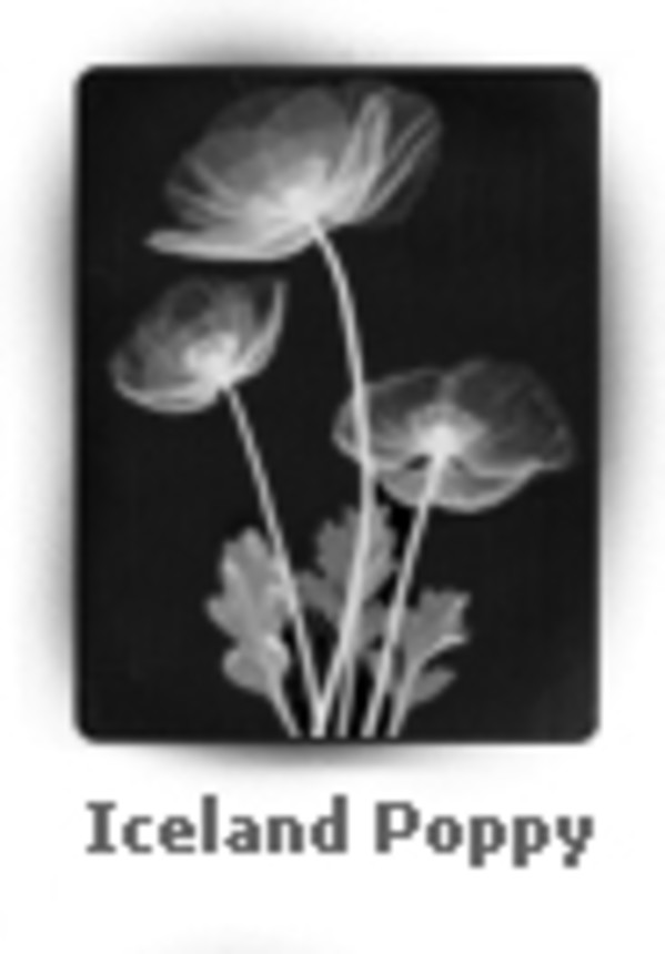
Dr. Bruwer was a longtime radiologist at TMC. In the 1960s he began experimenting with X-ray photography, utilizing a compact, lead-lined X-ray machine originally designed for hospital research. The low-voltage capability of the machine allowed Dr. Bruwer to capture the delicate structures of natural objects. He called these images skiagraphs: "skia" and "graph" come from the Greek words for “shadow drawing.” The three skiagraphs donated to the hospital by the artist's daughter are hanging just outside Radiology.
About the skiagraphs, from website:
Skiagraphics of plants and shells are sometimes mistaken for delicate pen-and-ink drawings or for black-and-white photographs. When you look more closely, it becomes clear you’re not seeing a two-dimensional rendering of nature’s beauty, you are actually seeing through the leaves and petals and into the shells’ chambers. That’s because skiagraphics are X-ray prints of natural objects. The word skiagraphic comes from the Greek skia meaning shadow and graphic referring to drawing. An X-ray print is actually composed of overlapping shadows. Whereas a photograph is created when light bounces off a subject and onto light sensitive film, an X-ray is created when X-rays pass through an object and create shadows on X-ray-sensitive film. When a doctor flips an X-ray of your lungs onto a light box, he is looking at overlapping shadows.