- Veronica Falconieri Hays
- Improving Resolution by Cryo-EM
- Color
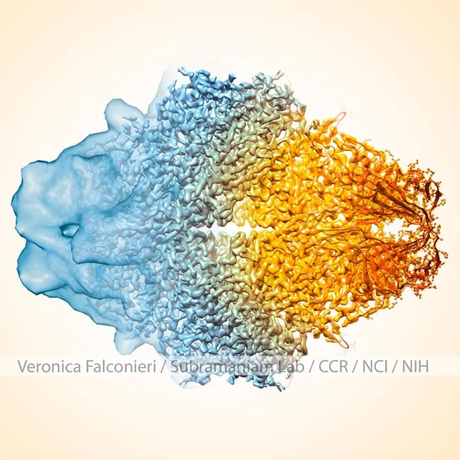
This molecule, beta-galactosidase, has been studied by the structural biology technique cryo-electron microscopy (cryoEM) in order to determine its structure. As cryo-EM technology has improved, the resolution of the resulting structures has improved from rough, low resolution maps (blue, left) to high resolution maps that enable model fitting (gold, right). Cryo-electron microscopy received the 2017 Nobel Prize in Chemistry, and is beginning to play an important role in drug development.
- Collections: Molecular
About the Artist: Veronica Falconieri Hays x
Veronica Falconieri Hays is a Certified Medical Illustrator in the Washington DC area specializing in medical, molecular, cellular, and biological visualization. She founded her company Falconieri Visuals in 2017 and works full time creating visuals for clients in biotech, pharma, and research fields.
Prior to founding Falconieri Visuals, Veronica earned her MA in Medical and Biological Illustration from Johns Hopkins University School of Medicine, where she studied medical subjects such as anatomy alongside medical students, as well as completing coursework in illustration and animation.
After graduating, Veronica worked within a cryo-electron microscopy lab at the National Cancer Institute. She collaborated extensively with researchers as they discovered structures of biological molecules, and is a published author in several journals including Science and Cell.
Education
MA, Medical & Biological Illustration, Johns Hopkins University School of Medicine.
BA, Biological Sciences, Smith College