- Carolina Hrejsa
- Eye Rotation on Visual-Motor Function in Frogs
- Adobe Illustrator, Adobe Photoshop
Collection: Herpetology x
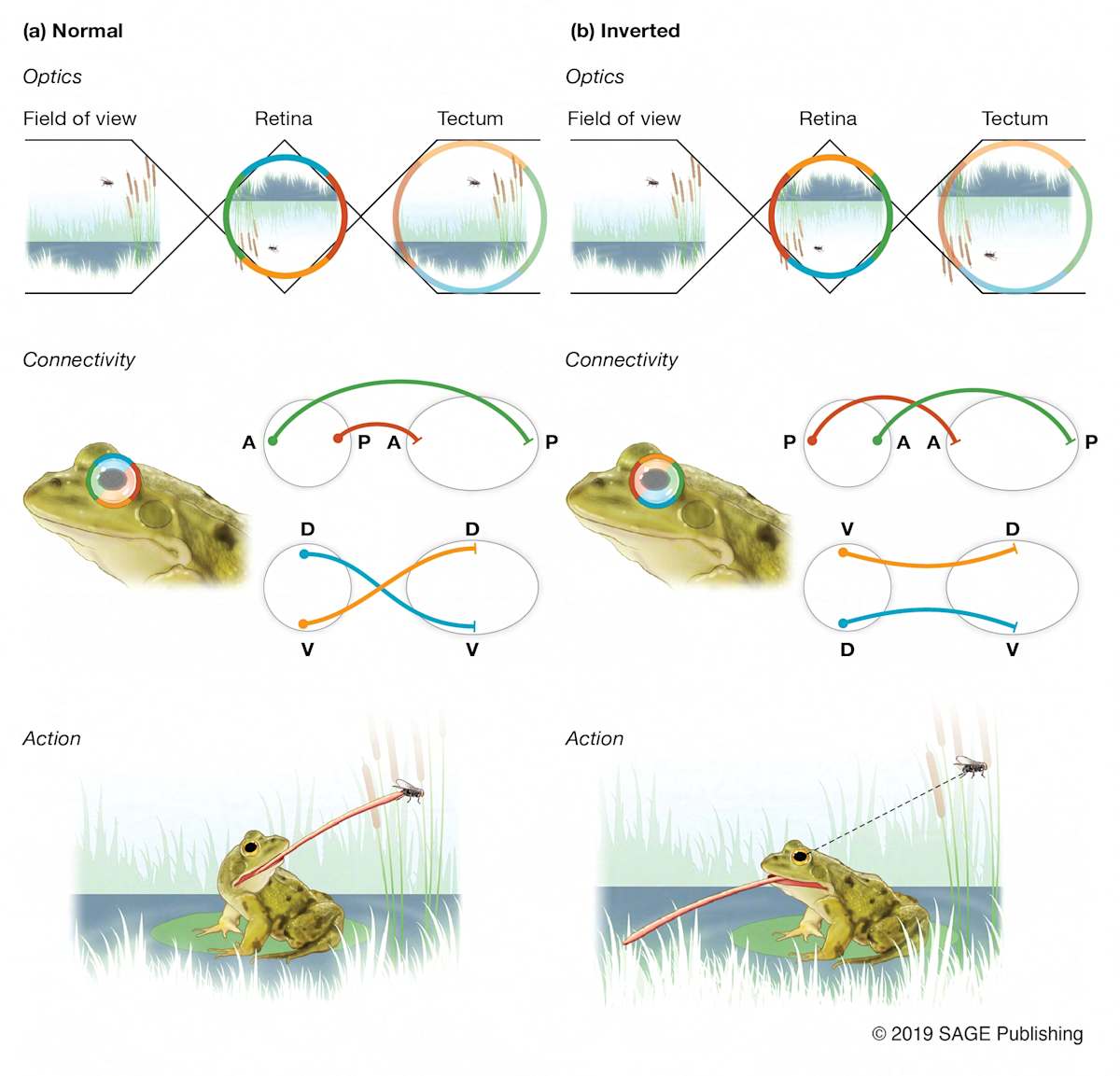
This figure illustrates Roger Sperry’s classic experiment on optic nerve regeneration and the chemoaffinity hypothesis. (a) In a normal frog, visual information from the retina is correctly mapped to the optic tectum, allowing accurate prey capture. (b) When the eye is surgically rotated 180 degrees and the optic nerve regenerates, the retinal-tectal connections are re-established based on their original anatomical coordinates rather than experience. As a result, the frog perceives the world as flipped and misdirects its strikes, supporting the idea that biochemical gradients guide neural connectivity.
Illustrated by Carol Hrejsa, CMI for Body Scientific International. © 2019 Sage Publishing
- Subject Matter: Animal Anatomy
- Collections: Herpetology
About the Artist: Carolina Hrejsa x
Carol Hrejsa, CMI, MS is a board-certified medical illustrator and biomedical visualization educator. She has over 15 years of experience creating, directing, and visualizing scientific and medical concepts. She earned her BA at the University of Chicago and studied with mentor Dr. Paul Sereno at the Dinosaur Fossil Lab. She then attended the UIC Biomedical Visualization program and graduated with an MS in medical illustration.
She worked as a lead illustrator and project manager at Body Scientific International for 16 years. Additionally, she has been an adjunct professor teaching Introduction to Science Visualization at the UIC life science visualization undergraduate program. Moreover, she runs her own studio, Hrejsa Medical Illustration. At the end of the day, she proudly declares herself a master of Adobe Photoshop, but her heart truly belongs to the art of traditional figure drawing.