- Carolina Hrejsa
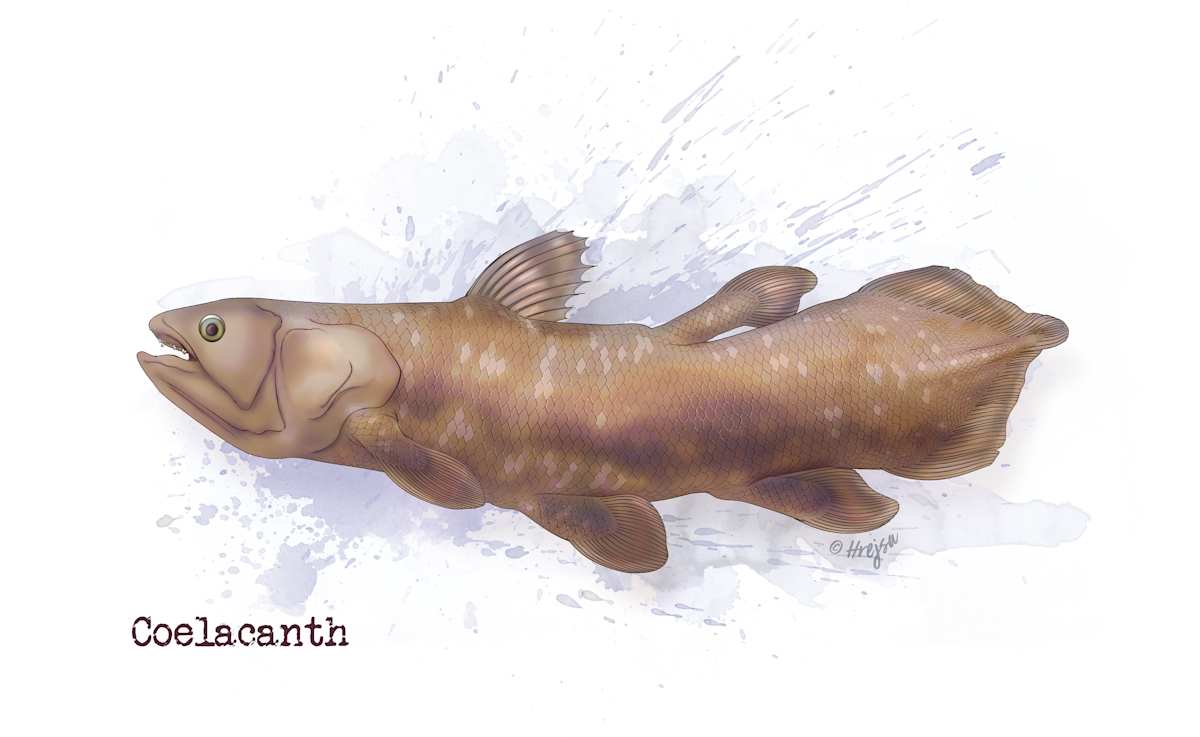
- Coelacanth
- Adobe Photoshop
This detailed illustration of the coelacanth, a living fossil, showcases its unique anatomical features. The image highlights the lobed pectoral and pelvic fins, which are crucial for its locomotion in deep-sea environments. The distinct three-lobed tail structure is also depicted, emphasizing its evolutionary significance. This image serves as an educational tool in marine biology and paleontology, aiding in the understanding of vertebrate evolution and anatomy.
- Subject Matter: Fish
- Collections: Aquatic Life
About the Artist: Carolina Hrejsa x
Carol Hrejsa, CMI, MS is a board-certified medical illustrator and biomedical visualization educator. She has over 15 years of experience creating, directing, and visualizing scientific and medical concepts. She earned her BA at the University of Chicago and studied with mentor Dr. Paul Sereno at the Dinosaur Fossil Lab. She then attended the UIC Biomedical Visualization program and graduated with an MS in medical illustration.
She worked as a lead illustrator and project manager at Body Scientific International for 16 years. Additionally, she has been an adjunct professor teaching Introduction to Science Visualization at the UIC life science visualization undergraduate program. Moreover, she runs her own studio, Hrejsa Medical Illustration. At the end of the day, she proudly declares herself a master of Adobe Photoshop, but her heart truly belongs to the art of traditional figure drawing.