- Veronica Falconieri Hays
- A Spitting Image in 3D
- Color
Collection: Molecular x
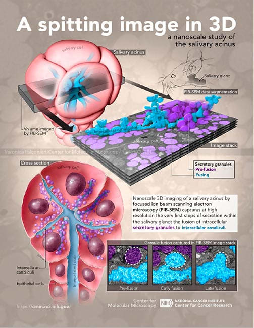
Subject: Nanoscopic 3D data of mouse salivary cells, salivary acinus anatomy Objective: Highlight the capabilities of focused ion beam scanning electron microscopy (FIB-SEM) in studying nanoscale biological features, using the example of a mouse salivary gland. These capabilities include not only 3D structural data, but many individual 2D images that capture multiple instances of a process, as demonstrated by the inset of the 3 stages of salivary vesicle fusion.
- Collections: Molecular
About the Artist: Veronica Falconieri Hays x
Veronica Falconieri Hays is a Certified Medical Illustrator in the Washington DC area specializing in medical, molecular, cellular, and biological visualization. She founded her company Falconieri Visuals in 2017 and works full time creating visuals for clients in biotech, pharma, and research fields.
Prior to founding Falconieri Visuals, Veronica earned her MA in Medical and Biological Illustration from Johns Hopkins University School of Medicine, where she studied medical subjects such as anatomy alongside medical students, as well as completing coursework in illustration and animation.
After graduating, Veronica worked within a cryo-electron microscopy lab at the National Cancer Institute. She collaborated extensively with researchers as they discovered structures of biological molecules, and is a published author in several journals including Science and Cell.
Education
MA, Medical & Biological Illustration, Johns Hopkins University School of Medicine.
BA, Biological Sciences, Smith College