Guild of Natural Science Illustrators
The GNSI is a 501(c)(3) that connects, provides professional development & networking opportunities to visual science communicators
Message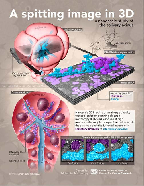
- Veronica Falconieri Hays
- A Spitting Image in 3D
- Color
Subject: Nanoscopic 3D data of mouse salivary cells, salivary acinus anatomy Objective: Highlight the capabilities of focused ion beam scanning electron microscopy (FIB-SEM) in studying nanoscale biological features, using the example of a mouse salivary gland. These capabilities include not only 3D structural data, but many individual 2D images that capture multiple instances of a process, as demonstrated by the inset of the 3 stages of salivary vesicle fusion.
- Collections: Molecular